The Knee
In human anatomy, the knee is the lower extremity joint connecting the femur and the tibia. Tthe knee supports nearly the entire weight of the body, and as a result it is prone to both acute injuries like ligament tears and chronic injuries such as osteoarthritis.
Function Of The Knee
The knee functions as a living, self-maintaining, biologic transmission, designed to accept and transfer biomechanical loads between the femur, tibia, patella, and fibula. In this analogy, the ligaments represent non-rigid adaptable sensate linkages, used to hold everything together within the biologic transmission. Articular cartilage acts as bearing surfaces, and the menisci as mobile bearings, smoothing out interactions between all the moving parts. The muscles function as living cellular engines that power movement in the joint as well as brakes and damping systems to absorb loads.
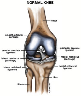
Knee Anatomy
The knee itself is actually comprised of two joints, known as the femoro-patellar and femoro-tibial joints.
The femoro-patellar joint consists of the patella, or “kneecap”, a sesamoid bone that sits within the tendon of the anterior (front) thigh muscle (m. quadriceps femoris), and the patellar groove on the front of the femur through which it slides.
The femoro-tibial joint links the femur, or thigh bone, with the tibia, the main bone of the (lower) leg. The joint is bathed in a viscous (synovial) fluid which is contained inside the “synovial” membrane, or joint capsule. The recess behind the knee is called the popliteal fossa.